Technology
Cone Beam Computed Tomography
What is Cone Beam Computed Tomography (CBCT)?

CBCT is an advanced, medical imaging technique that produces three dimensional images in contrast to the traditional, two dimensional images produced by conventional dental radiographs. The clarity and dimension of the images significantly enhances the ability to see the positioning and spatial relationship of the teeth, jaw bone and other vital structures such as sinuses and large nerve bundles. CBCT imaging also provides cross section views of teeth not possible using conventional two dimensional x-rays. The imaging is an integration of medical CT scan technology and dental panoramic imaging. In cone beam computed tomography imaging, the machine is sending the X-rays in a divergent cone-shape.
What Can Patients Expect?
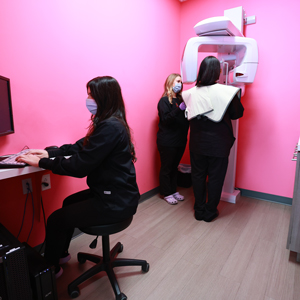
For patients, the process of obtaining CBCT images will seem remarkably similar to having panoramic x-rays taken. A scanner will rotate around your head for approximately 15 seconds. The machine is open and does not feel uncomfortably enclosed. Undergoing a CBCT scan requires little to no special preparation, and no pain or discomfort is expected. Metal objects, eyeglasses and hairpins may affect the images and should be removed prior to your scan. You may also be asked to remove hearing aids and/or any removable dental prosthesis.
Do we replace conventional X-Rays with CBCT?
No. Using CBCT is not our first course of action. We will continue to use conventional, digital x-rays as the initial step in your examination and as we decide on a plan of action for your endodontic care. Only in certain cases where regular dental x-rays are not sufficient will we recommend follow-up with CBCT imaging to aid in diagnosis.
